
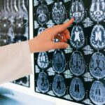
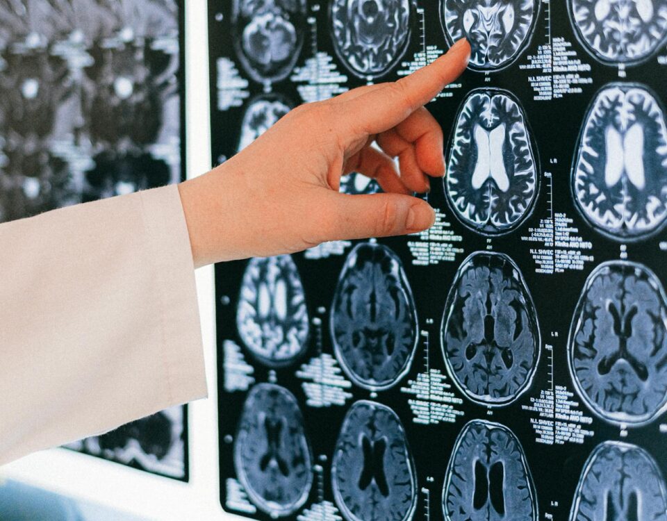
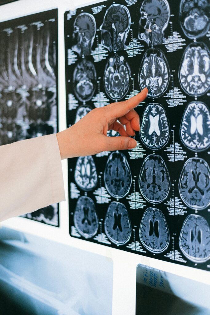
Zombie Vasculature: Endothelial Senescence, Long COVID & Senolytics — The Vascular Reset Protocol
April 21, 2026
Urolithin A Mitophagy: The Post-Viral Immune Recovery Protocol Backed by 2025 RCT Data
June 15, 2026
EBV reactivation Long COVID microaggregates represent a compounding, dual-hit mechanism that may explain why a significant subset of Long COVID patients experience fatigue that standard care simply cannot resolve. If you’re a finance or banking professional who “recovered” from COVID-19 but is still running at 60% capacity months later — fighting brain fog, post-exertional crashes, and cardiovascular instability — there’s now emerging mechanistic evidence pointing directly at this two-vector biological failure.
What this means in plain terms: Your SARS-CoV-2 infection may have flipped a switch on a dormant virus you’ve carried for decades, while simultaneously seeding your bloodstream with clots too small to show up on standard imaging but large enough to starve your tissues of oxygen.
Executive Summary: The Alpha of the Dual-Hit Protocol
- Hit 1 — Viral Reactivation: SARS-CoV-2 infection destabilizes EBV latency in B cells, triggering a secondary immune cascade that drives mitochondrial fragmentation, sustained inflammation, and the Systemic Pro-inflammatory Secretory Phenotype (SASP) associated with cellular senescence.
- Hit 2 — Vascular Obstruction: The SARS-CoV-2 spike protein nucleates fibrinogen into amyloid-like microaggregates resistant to normal fibrinolysis. These microscopic clots obstruct capillary flow, creating tissue-level hypoxia that drives ATP deficits and Post-Exertional Malaise (PEM).
- The Evidence: A 2026 Scientific Reports study (Wick et al.) confirmed both mechanisms co-occur in 80% of Long COVID patients with positive EBV T-cell responses, and demonstrated 78% self-assessed clinical improvement with combined anti-thrombotic + valaciclovir therapy versus only 38% with anti-thrombotic treatment alone.
The Biological Mechanism: How EBV Reactivation and Long COVID Microaggregates Create a Vicious Cycle
Step 1: SARS-CoV-2 Unlocks Latent EBV
Approximately 95% of adults carry Epstein-Barr Virus in a latent state, silenced within B lymphocytes through epigenetic hypermethylation. Under normal conditions, a competent T-cell surveillance system maintains this dormancy. SARS-CoV-2 disrupts this equilibrium.
Research published in Cell Host & Microbe and supported by immunological data from a 2022 NIH-indexed cohort study found that Long COVID symptom burden correlated with serological evidence of recent EBV reactivation — including elevated IgG antibodies to Early Antigen (EA), a specific marker of lytic reactivation rather than past exposure.
The proposed mechanism centers on EBNA2 and viral dUTPase proteins. Professor Bhupesh Prusty at the University of Würzburg has shown that EBV dUTPase protein, secreted during early lytic reactivation, directly interferes with mitochondrial dynamics — specifically the fusion-fission cycle governed by DRP1 and MFN2. The result is pathological mitochondrial fragmentation: smaller, less efficient mitochondria unable to produce adequate ATP via oxidative phosphorylation. This is bioenergetic failure at the organelle level.
What this means for you: Your cells are running on a dying battery. The CPU clock speed has dropped because the power supply is compromised — not because you’re lazy or “deconditioned.”
Step 2: The Microaggregate Formation Cascade
Simultaneously, the SARS-CoV-2 spike protein drives a separate vascular catastrophe. Research by Prof. Resia Pretorius at Stellenbosch University, most comprehensively documented in a landmark 2022 Cardiovascular Diabetology study (PMID 35549427), demonstrated that fibrinogen in Long COVID blood samples undergoes amyloid-like misfolding when exposed to circulating spike protein or LPS. The resulting microclots:
- Resist normal plasmin-mediated fibrinolysis (the body’s clot-clearing system)
- Range from 1–200 μm in diameter — large enough to obstruct terminal capillaries
- Entrap pro-inflammatory proteins including α2-antiplasmin, SAA, fibronectin, and Von Willebrand Factor
- Persist for months to years post-acute infection
The confirmatory Wick et al. Scientific Reports (2026) study extended this work by directly visualizing circulating microaggregates in peripheral venous blood via live confocal fluorescence microscopy — confirming structures of 100–200 μm containing leukocytes, granulocytes, thrombocytes, and unidentified carbohydrate-rich material. These aggregates were stable overnight and demonstrated adhesive capacity consistent with active hemostatic involvement.
Step 3: The Compounding Feedback Loop
Here’s where EBV reactivation Long COVID microaggregates become particularly insidious as a combined pathology:
- EBV-reactivated cells activate the SASP — secreting IL-6, TNF-α, and MMP-3 into the vascular environment
- This inflammatory milieu further destabilizes endothelial function, amplifying platelet activation and aggregate formation
- Microaggregates obstruct capillary perfusion → tissue hypoxia → reduced oxygen delivery to mitochondria
- Hypoxic mitochondria shift from oxidative phosphorylation to anaerobic glycolysis (the “Warburg-like” metabolic reprogramming observed in ME/CFS)
- ATP deficit triggers redox imbalance (excess ROS production) which further damages mitochondrial cristae membranes
- Cristae swelling impairs ATP synthase function — completing a devastating feedback loop of compounding bioenergetic failure
This self-amplifying cycle is why 45% of COVID-19 survivors report unresolved symptoms at four-month follow-up, and why mitochondrial health interventions alone prove insufficient when the vascular obstruction component remains unaddressed.
The Interventions: A 4-Layer Protocol Stack for EBV Reactivation and Long COVID Microaggregates
Standard care — rest, graded exercise therapy (GET), and generic anti-inflammatories — addresses none of the four core mechanisms. The emerging evidence supports a mechanistically targeted four-layer approach. Each layer addresses a distinct failure point in the cascade.
Layer 1: Antiviral Targeting (EBV Suppression)
Agent: Valaciclovir (1,500–3,000 mg/day in divided doses)
The Wick et al. (2026) cohort demonstrated that patients receiving both valaciclovir and anti-thrombotic therapy achieved a Bell Score improvement of +43 points — nearly double the +23 points seen with anti-thrombotic therapy alone. The 78% subjective improvement rate is clinically significant, though the study’s small sample (n=5–16 per group) warrants cautious interpretation. The Yale LISTEN trial is currently evaluating antiviral approaches in Long COVID in a larger, controlled design.
Note: Valaciclovir specifically targets herpesvirus polymerase. EBV is only moderately sensitive; clinical benefit may also derive from suppression of co-reactivating HHV-6A or HSV-1, which Prusty’s research implicates in the same mitochondrial fragmentation pathway.
Layer 2: Fibrinolytic Support (Microaggregate Dissolution)
Agents: Nattokinase (2,000–4,000 FU/day) and/or Lumbrokinase
Both are serine proteases that degrade fibrin and fibrin-derived microclots via pathways distinct from endogenous plasmin. Critically, they appear effective against the amyloid-form fibrin that resists normal fibrinolysis. For a detailed mechanistic breakdown, see our dedicated analysis of nattokinase in Long COVID microclots. The Wick et al. data complement this by demonstrating that anti-platelet therapy (aspirin 100 mg + low molecular weight heparin) reduced aggregate counts measurably — with the ASS + heparin combination outperforming clopidogrel in social reintegration (78% vs. 33%).
Layer 3: Mito-Resuscitation Stack
Agents: CoQ10 (300–600 mg/day ubiquinol form), NMN or NR (500–1,000 mg/day NAD+ precursors), PQQ (20 mg/day)
The goal here is to restore electron transport chain efficiency in mitochondria operating under the combined stress of EBV-driven fragmentation and hypoxia-induced redox imbalance. CoQ10 functions as an essential electron carrier at Complex I–III; depletion is documented in ME/CFS and Long COVID. NAD+ precursors support SIRT1/SIRT3 activity (critical for mitochondrial quality control via mitophagy). PQQ stimulates PGC-1α — the master regulator of mitochondrial biogenesis — effectively prompting the cell to build new mitochondria to replace dysfunctional ones.
For a full discussion of this protocol in the context of high-performance professionals, see our mitochondrial optimization guide.
Layer 4: SASP Suppression and Neuroinflammation Dampening
Agents: Low-Dose Naltrexone (LDN, 1.5–4.5 mg/night), Quercetin (1,000–2,000 mg/day), Omega-3 EPA/DHA (3–4 g/day EPA-dominant)
LDN’s mechanism in post-viral states operates via transient μ-opioid receptor blockade, upregulating endogenous opioid production and — critically — antagonizing TLR4 microglial activation. Microglial activation drives the neuroinflammatory component of brain fog and cognitive dysfunction in Long COVID. Our deep-dive into LDN and TRPM3 channel dysfunction in Long COVID covers the neurological mechanisms in detail.
Quercetin provides dual function: as a senolytic (clearing EBV-primed senescent cells expressing SASP) and as a mild fibrinolytic complement to Layers 2 and 3. For context on how senescent endothelial cells amplify vascular dysfunction in this setting, see our analysis of endothelial senescence and senolytics in Long COVID.
Standard Care vs. Emerging Dual-Hit Protocol: EBV Reactivation and Long COVID Microaggregates
| Parameter | Standard Care | Emerging Dual-Hit Protocol |
|---|---|---|
| EBV reactivation assessment | Not routinely tested; EBV serology only if mononucleosis suspected | EliSpot T-cell reactivity to EBV peptides; sensitive early-reactivation detection |
| Microaggregate detection | Not tested; absent from standard coagulation panels | Live confocal fluorescence microscopy of native blood; cytopathology protocol |
| Antiviral component | None (unless active lytic EBV confirmed by PCR) | Valaciclovir 1,500–3,000 mg/day targeting herpesvirus family |
| Vascular/fibrinolytic support | Not indicated without DVT/PE diagnosis | Aspirin + LMWH (anti-platelet) + nattokinase/lumbrokinase (fibrinolytic) |
| Mitochondrial support | Not addressed; fatigue attributed to deconditioning | CoQ10 + NAD+ precursors + PQQ mito-resuscitation stack |
| Senolytic/SASP suppression | Not indicated in standard Long COVID protocols | Quercetin + Fisetin for EBV-primed senescent cell clearance |
| Neuroinflammation | CBT, pacing (no pharmacological target) | Low-Dose Naltrexone (LDN) for microglial dampening; Omega-3 for resolution |
| Outcome (self-assessed improvement) | 38% (anti-thrombotic monotherapy; Wick et al. 2026) | 78% (combined anti-thrombotic + virostatic; Wick et al. 2026) |
| Social reintegration rate | 33% (clopidogrel arm; Wick et al. 2026) | 78% (ASS + heparin + valaciclovir arm; Wick et al. 2026) |
| Evidence base | Guideline-based; symptom management only | Emerging (2024–2026); mechanistic; early pilot data promising |
Executive Takeaways: Re-Optimizing, Not “Recovering”
In our clinical review of the emerging literature, the framing of “Long COVID recovery” consistently understates the challenge. This is not a linear return to baseline. For the finance professional, this is a system re-architecture problem: your bioenergetic infrastructure has taken damage from two concurrent vectors, and patching one without addressing the other produces partial and often temporary improvement.
The actionable framework:
- Get the right diagnostics first. Standard blood work will miss both EBV reactivation (you need EliSpot or early antigen IgG, not just IgG to VCA) and microaggregates (requires specialist confocal microscopy — not a standard coagulation panel). Without proper phenotyping, you may be treating the wrong target.
- Respect the PEM threshold — rigorously. Graded exercise therapy in the presence of active microaggregates and bioenergetic failure is contraindicated. Pushing through PEM in this state causes measurable biological harm. Treat your energy envelope as a hard capital constraint, not a psychological barrier.
- Layer the protocol sequentially. The data suggests anti-thrombotic + antiviral first, then mito-resuscitation support, then SASP management. Attempting all simultaneously creates confounding variables and increases adverse event risk.
- Retest at 3–6 months. Bell Score trajectory, microaggregate counts, and EBV T-cell reactivity are the KPIs. If you’re not tracking, you’re not managing.
For professionals experiencing dysautonomia symptoms alongside fatigue — heart rate variability instability, POTS-like episodes, blood pressure dysregulation — consider reviewing the evidence on Stellate Ganglion Block as a neuromodulatory intervention for the autonomic dysfunction component of Long COVID.
Conclusion
The compounding relationship between EBV reactivation, Long COVID microaggregates, and bioenergetic failure represents one of the most mechanistically coherent explanations for why a subset of Long COVID patients remain functionally impaired despite resolution of acute infection. The Wick et al. (2026) data, while preliminary, provides the first direct clinical evidence that targeting both vectors simultaneously produces meaningfully superior outcomes to monotherapy. This is not a “cure” narrative — it is a mechanistic signal that the field is beginning to operationalize into testable, tractable interventions.
The finance and banking professional managing post-viral impairment has more analytical leverage here than most patients: the ability to read primary data, quantify symptom trajectories, and pressure-test clinical decisions against emerging evidence. That’s not a small advantage in a disease where the standard-of-care is still years behind the science.
Medical Disclaimer: The information in this article is intended for educational purposes only and does not constitute medical advice. The interventions discussed — including valaciclovir, low-dose naltrexone, nattokinase, and other pharmacological or nutraceutical agents — should only be undertaken under the supervision of a qualified medical professional. Do not modify any medication regimen without consulting your physician. This article does not establish a doctor-patient relationship. Always seek professional medical evaluation before pursuing any treatment described here.

